
How Do Peptides Work? Understanding Receptors and Biological Activity
Exploring peptide-receptor interactions, cellular signaling pathways, molecular recognition, and biological responses
Peptides participate in numerous biological processes by interacting with specific molecular targets within living systems. Their activities are closely connected to the way their molecular structures are recognized by receptors, enzymes, and other biological components.
In cellular biology, these interactions are particularly important because they allow cells to receive and respond to chemical signals. Certain peptides function as hormones, neurotransmitters, or signaling molecules, helping regulate processes such as metabolism, immune activity, cellular communication, and physiological coordination.
But how does a peptide actually produce a biological response?
The answer involves several interconnected mechanisms, including molecular recognition, receptor binding, conformational changes, signal transduction, and downstream cellular activity.
Understanding these mechanisms is essential in modern molecular biology, biochemistry, pharmacology, and biotechnology.
This guide examines how peptides interact with biological receptors, how signals are transmitted within cells, and which molecular factors influence peptide activity.
1. How Do Peptides Work in Biological Systems?
Peptides work through interactions with biological molecules that recognize and respond to their chemical structures.
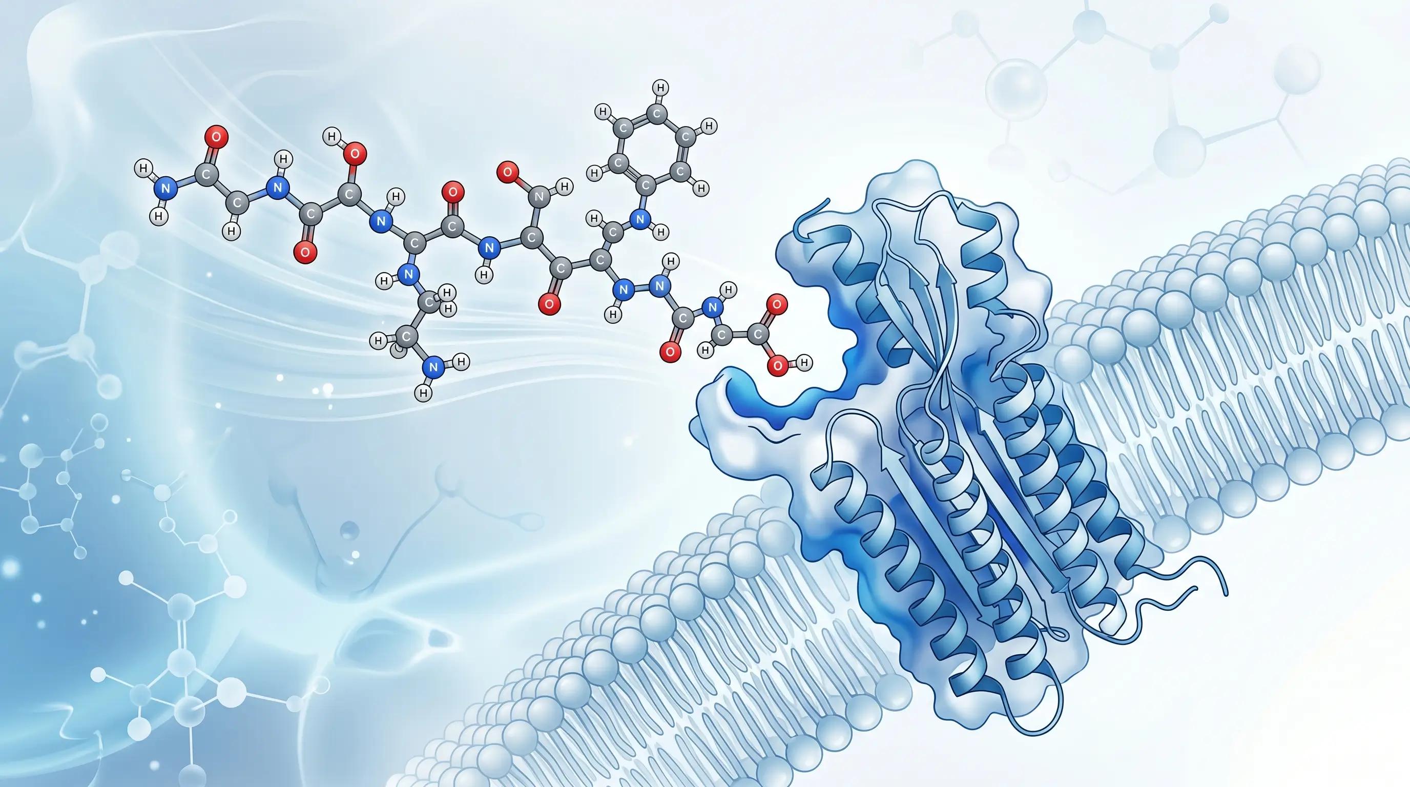
Many biologically active peptides function as ligands. A ligand is a molecule that binds to a specific biological target, such as a receptor, enzyme, or other protein.
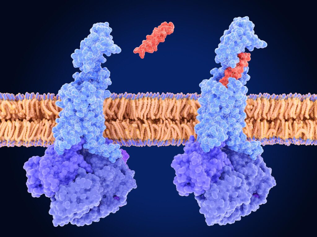
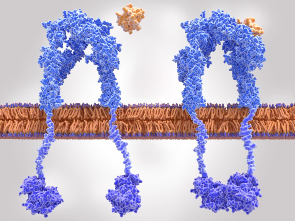
When a peptide binds to a compatible receptor, it can alter the receptor’s molecular conformation and influence its activity.
In many cases, this interaction initiates a sequence of intracellular events known as signal transduction.
Signal transduction is the process through which a cell converts an external or internal signal into a biological response.
Depending on the peptide and its target, the resulting response may involve changes in enzyme activity, ion movement, cellular metabolism, secretion, gene expression, or other physiological functions.
Not all peptides operate through the same mechanism. Their effects depend on their molecular properties, receptor type, target-cell characteristics, and the biological environment.
2. What Are Peptide Receptors?
Peptide receptors are biological molecules, usually proteins, that recognize and bind particular peptide ligands.
Many peptide receptors are located on the surface of cell membranes. This arrangement is particularly important because many peptide hormones are hydrophilic and cannot freely diffuse through the lipid bilayer of the plasma membrane.
Instead, they interact with receptors exposed on the extracellular surface of target cells.
Receptor recognition depends on several molecular characteristics, including:
- The amino acid sequence of the peptide.
- The peptide’s three-dimensional conformation.
- The chemical properties of its side chains.
- The structure and binding characteristics of the receptor.
- The surrounding biological environment.
A peptide may bind strongly to one receptor but interact weakly or not at all with another.
This molecular selectivity contributes to the specificity of biological signaling.
Receptor Recognition and Molecular Compatibility
Peptide-receptor recognition involves complementary chemical and structural interactions.
These may include hydrogen bonding, electrostatic interactions, hydrophobic interactions, and other non-covalent forces.
The binding process can stabilize particular conformations of the receptor, influencing whether and how the receptor activates downstream signaling.
It is important to understand that receptor binding does not automatically mean that a biological response will occur. A molecule may bind to a receptor without activating it.
The functional outcome depends on the nature of the ligand-receptor interaction.
3. How Does Peptide-Receptor Binding Produce a Cellular Response?
Peptide signaling can be understood through a series of connected molecular events.
Step 1: Peptide Recognition
A peptide reaches a target cell through a biological signaling route. Depending on the system, it may be released locally or transported through the bloodstream.
The peptide encounters cells that express compatible receptors.
Step 2: Receptor Binding
The peptide interacts with a suitable binding region on the receptor.
This interaction is influenced by the molecular structure of both the ligand and the receptor.
Step 3: Receptor Conformational Change
In many receptor systems, ligand binding changes the receptor’s conformational state.
This structural rearrangement can expose or activate intracellular regions involved in signal transmission.
Step 4: Intracellular Signal Transduction
The activated receptor influences intracellular signaling components.
These may include G proteins, protein kinases, enzymes, ion channels, or other regulatory molecules.
Step 5: Cellular Response
The signaling cascade eventually influences cellular activity.
Depending on the biological system, the response may involve changes in metabolism, secretion, ion transport, gene expression, or other cellular processes.
Step 6: Signal Regulation
Cellular signaling is controlled through mechanisms that limit, modify, or terminate receptor activity.
These mechanisms help prevent excessive or prolonged stimulation and allow cells to respond appropriately to changing biological conditions.
This sequence provides a simplified model. Actual receptor signaling may involve multiple pathways operating simultaneously.
4. Major Types of Receptors Involved in Peptide Signaling
Peptides can interact with several classes of receptors. Understanding these receptor families helps explain why different peptides can produce different biological effects.
4.1 G-Protein-Coupled Receptors (GPCRs)
G-protein-coupled receptors are among the most important receptor families involved in peptide signaling.
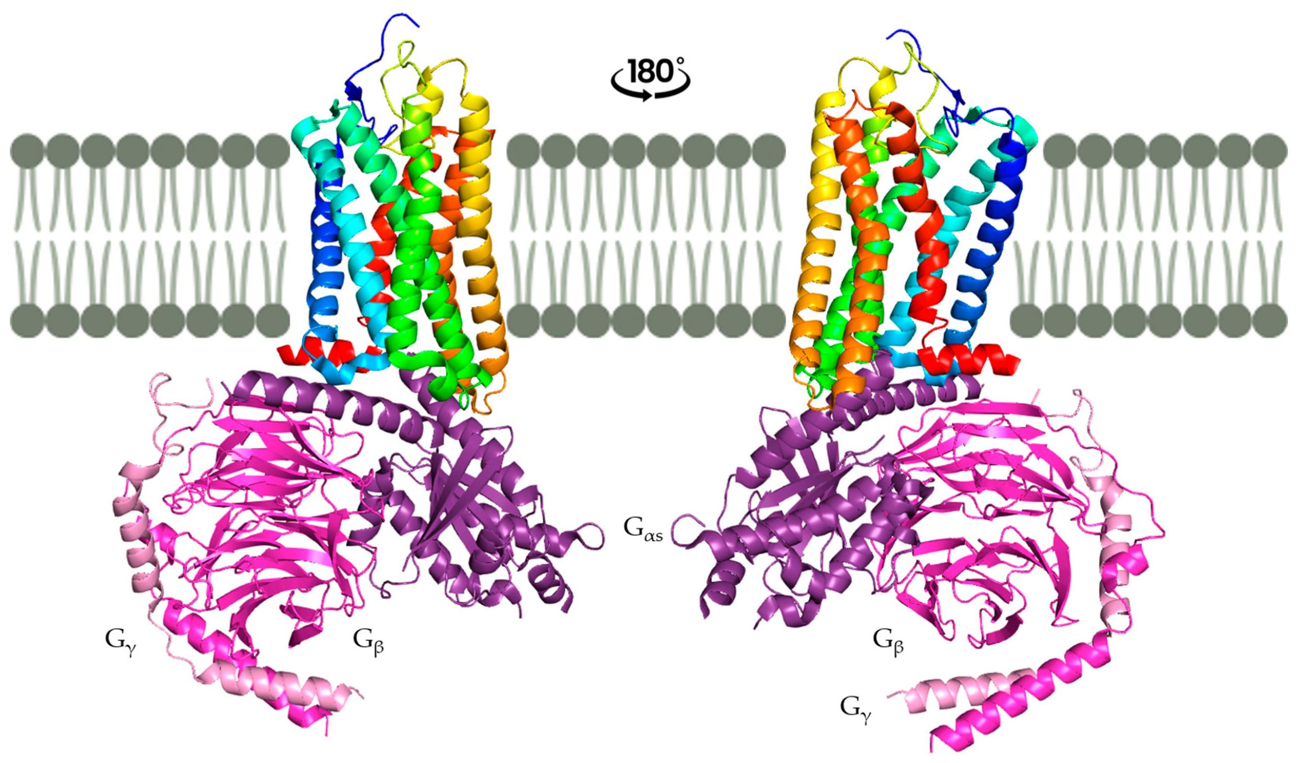
GPCRs are membrane proteins characterized by seven transmembrane alpha-helical segments.
They recognize different extracellular signals, including peptide hormones, neuropeptides, neurotransmitters, and other biological molecules.
When a compatible peptide binds to a GPCR, the receptor undergoes a conformational change that allows it to activate an associated heterotrimeric G protein.
The G protein contains three subunits:
- Alpha (α).
- Beta (β).
- Gamma (γ).
In its inactive state, the alpha subunit is associated with guanosine diphosphate (GDP).
Receptor activation promotes the exchange of GDP for guanosine triphosphate (GTP), allowing the G protein to regulate downstream signaling components.
Different G-protein families activate different intracellular pathways.
4.2 Receptor Tyrosine Kinases (RTKs)
Receptor tyrosine kinases are another important class of membrane-associated receptors.
These receptors contain intracellular regions with tyrosine kinase activity.
Insulin provides an important example of a peptide hormone that acts through a receptor tyrosine kinase.
When insulin binds to its receptor, the receptor undergoes structural changes that activate its intracellular kinase activity.
This leads to phosphorylation of specific tyrosine residues and recruitment or activation of downstream signaling proteins.
Two important pathways associated with insulin receptor signaling are:
- The PI3K-AKT pathway, which contributes to metabolic regulation and other cellular processes.
- The Ras-MAPK pathway, which participates in cellular growth, differentiation, and related responses.
These pathways illustrate how a peptide hormone can influence intracellular processes through a receptor that does not belong to the GPCR family.
4.3 Cytokine Receptors and JAK-STAT Signaling
Some peptide or protein signaling molecules interact with cytokine receptor systems.
These receptors do not possess intrinsic tyrosine kinase activity in the same way as receptor tyrosine kinases.
Instead, they associate with intracellular Janus kinases (JAKs).
When an appropriate ligand binds, receptor rearrangement or association brings the relevant JAK molecules into an activated configuration.
JAKs phosphorylate receptor regions and facilitate the recruitment and phosphorylation of signal transducers and activators of transcription (STATs).
Activated STAT proteins can then move into the nucleus and influence gene transcription.
This pathway is important in various immune, growth, and hematopoietic signaling processes.
Not every cytokine is a short peptide, and cytokine receptors should not be treated as a single universal peptide receptor mechanism.
5. Understanding Second Messengers in Peptide Signaling
Many peptide-receptor interactions trigger intracellular signaling through molecules known as second messengers.
A second messenger is an intracellular molecule or ion that helps transmit and amplify information from an activated receptor.
These molecules are important because a relatively small number of activated receptors can influence multiple downstream cellular components.
Some of the best-known second messengers include:
Cyclic Adenosine Monophosphate (cAMP)
cAMP is generated from ATP by the enzyme adenylyl cyclase.
When certain GPCRs activate stimulatory G proteins, adenylyl cyclase activity increases, resulting in higher intracellular cAMP concentrations.
cAMP can activate protein kinase A (PKA), which phosphorylates target proteins and modifies their activity.
This pathway is involved in several hormonal and metabolic signaling processes.
Calcium Ions (Ca²⁺)
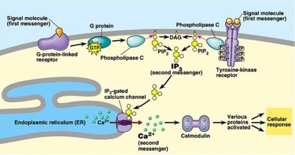
Calcium ions function as important intracellular signaling molecules.
Some receptor pathways promote calcium release from intracellular stores or calcium entry across the plasma membrane.
Changes in intracellular calcium concentrations can influence secretion, muscle contraction, enzyme activity, and other cellular functions.
Inositol Triphosphate (IP3)
IP3 is produced through the activity of phospholipase C, which cleaves a membrane phospholipid known as phosphatidylinositol 4,5-bisphosphate (PIP2).
IP3 can bind to receptors on the endoplasmic reticulum and promote the release of stored calcium into the cytoplasm.
Diacylglycerol (DAG)
DAG is generated alongside IP3 during PIP2 cleavage.
It remains associated with the plasma membrane and participates in the activation of protein kinase C (PKC), often in cooperation with calcium.
Together, these signaling components help cells translate receptor activation into specific biochemical responses.
6. Examples of Peptide Signaling in Biological Systems
Different peptides interact with different receptor systems, illustrating the diversity of biological signaling.
Insulin and Metabolic Signaling
Insulin is a peptide hormone produced by pancreatic beta cells.
It binds to the insulin receptor, a receptor tyrosine kinase, and initiates intracellular signaling pathways.
These pathways contribute to glucose uptake in responsive tissues, glycogen synthesis, lipid metabolism, and other metabolic activities.
Insulin is an important example of how peptide-receptor interactions influence whole-body physiology.
Glucagon and Glucose Regulation
Glucagon is a peptide hormone produced by pancreatic alpha cells.
It binds to glucagon receptors, which are GPCRs expressed prominently in the liver.
Receptor activation stimulates intracellular signaling involving Gs proteins, adenylyl cyclase, and cAMP.
The resulting signaling contributes to the regulation of hepatic glucose production and metabolism.
Oxytocin and Cellular Signaling
Oxytocin is a peptide hormone that interacts with the oxytocin receptor, a GPCR.
Receptor activation can stimulate Gq-associated signaling, including phospholipase C activity and intracellular calcium mobilization.
These mechanisms contribute to physiological processes such as uterine contraction and milk ejection.
Glucagon-Like Peptide-1 (GLP-1)
GLP-1 is an incretin peptide involved in glucose-dependent regulation of insulin secretion.
It interacts with the GLP-1 receptor, a class B GPCR.
Activation of this receptor stimulates intracellular signaling that includes cAMP-dependent pathways.
GLP-1 receptor signaling is an important subject in metabolic research and pharmaceutical development.
These examples demonstrate that the biological effect of a peptide is determined not simply by its presence, but by its interaction with a specific receptor and the signaling machinery available in the target cell.
7. What Determines the Biological Activity of a Peptide?
A peptide’s biological activity depends on several molecular and physiological factors.
Amino Acid Sequence
The amino acid sequence influences the chemical properties and molecular recognition characteristics of a peptide.
Changes in sequence may affect receptor binding, molecular stability, and biological activity.
Three-Dimensional Conformation
The spatial arrangement of a peptide influences how it interacts with its target.
Some peptides adopt specific conformations that are important for receptor recognition.
Receptor Affinity
Affinity describes the strength of the interaction between a ligand and its receptor under defined experimental conditions.
It is commonly evaluated through binding measurements, including the equilibrium dissociation constant (Kd).
A lower Kd generally indicates higher binding affinity.
However, affinity alone does not establish how strongly a peptide activates a receptor.
Receptor Efficacy
Efficacy refers to the ability of a ligand to produce a functional response through receptor activation.
A peptide may bind to a receptor but produce little or no activation.
This distinction is important when comparing receptor ligands.
Potency
Potency describes the concentration of a ligand required to produce a specified level of biological response.
In functional assays, potency is commonly expressed through the half-maximal effective concentration (EC50).
Potency is influenced by several factors, including affinity, efficacy, receptor abundance, and the characteristics of the experimental system.
Molecular Stability
A peptide’s stability can influence how long it remains available to interact with its biological target.
Some peptides are susceptible to enzymatic degradation by proteases and peptidases.
Their chemical structure and surrounding biological environment can influence their susceptibility to degradation.
Receptor Distribution
A peptide can only produce a direct receptor-mediated effect in cells that express the relevant receptor and possess the necessary signaling machinery.
Differences in receptor expression between tissues can therefore influence biological responses.
8. How Do Cells Regulate Peptide Signaling?
Cellular signaling must be carefully regulated to maintain biological balance.
Continuous receptor activation can result in changes in receptor responsiveness and the intensity of downstream signaling.
Several mechanisms help regulate these processes.
Receptor Desensitization
Desensitization occurs when a receptor becomes less responsive despite the continued presence of a signaling molecule.
In GPCR systems, receptor phosphorylation and the recruitment of regulatory proteins such as beta-arrestins can reduce receptor coupling to G proteins.
Receptor Internalization
Some activated receptors are removed temporarily from the cell surface through endocytosis.
This process can influence receptor availability and signaling duration.
Internalized receptors may be recycled back to the membrane or directed toward degradation, depending on the receptor and cellular context.
Receptor Downregulation
Downregulation involves a reduction in the number of receptors available within a cell.
It may occur through changes in receptor synthesis, degradation, or trafficking.
Signal Termination
Signaling pathways also contain mechanisms that switch off intracellular components.
Examples include GTP hydrolysis by G proteins, degradation of second messengers, and dephosphorylation of signaling proteins by phosphatases.
These regulatory mechanisms allow cells to control the timing, strength, and duration of biological responses.
9. How Do Scientists Study Peptide-Receptor Interactions?
Understanding peptide activity requires more than identifying a peptide sequence.
Researchers use several experimental methods to investigate receptor binding, molecular recognition, and downstream biological responses.
Receptor Binding Assays: These experiments investigate whether a peptide interacts with a particular receptor and help estimate binding affinity.
Functional Cell-Based Assays: Researchers measure changes in cellular activity after exposure to a peptide. Depending on the receptor, measurements may include cAMP production, calcium mobilization, or other signaling responses.
Surface Plasmon Resonance (SPR): SPR can be used to investigate molecular binding interactions and determine association and dissociation characteristics under controlled conditions.
Fluorescence-Based Assays: Fluorescent methods can help researchers monitor receptor activation, molecular interactions, or intracellular signaling.
Western Blotting: This technique can be used to investigate changes in the phosphorylation or abundance of specific signaling proteins.
Structural Biology: Cryo-electron microscopy, X-ray crystallography, and other structural techniques help scientists investigate the molecular arrangements of receptor-ligand complexes.
Combining binding, functional, and structural methods provides a more complete understanding of peptide activity than relying on a single measurement.
10. Why Is Peptide Signaling Important in Biotechnology?
Peptide signaling is an important research area because it connects molecular recognition with biological function.
Understanding these pathways contributes to several fields of biotechnology.
Drug Discovery
Researchers investigate peptide-receptor interactions to identify potential therapeutic candidates and understand how molecular changes influence receptor activity.
Metabolic Research
Peptide hormones such as insulin, glucagon, and GLP-1 are important subjects in research involving metabolic regulation and glucose homeostasis.
Neuroscience
Neuropeptides interact with specific receptors and contribute to the regulation of neural communication and physiological processes.
Immunological Research
Peptide and cytokine signaling systems participate in immune-cell communication, inflammation, and the regulation of immune responses.
Molecular Engineering
Scientists investigate how changes in peptide sequences and structures influence receptor recognition, stability, and signaling properties.
These applications demonstrate why receptor pharmacology and peptide signaling remain important subjects in modern biotechnology.
11. Future Directions in Peptide Signaling Research
Advances in structural biology, molecular modeling, computational chemistry, and biotechnology continue to expand scientific understanding of peptide-receptor interactions.
Several research directions are particularly important:
- Structural receptor analysis: Improved imaging and structural methods are helping researchers examine peptide-bound receptor conformations.
- Biased signaling: Scientists are investigating how different ligands can influence distinct signaling pathways through the same receptor.
- Computational peptide design: Computational tools are being explored to predict molecular interactions and guide peptide discovery.
- Advanced receptor assays: New analytical platforms are helping researchers investigate binding, signaling, and receptor regulation with greater detail.
- Personalized molecular research: Differences in receptor expression and biological pathways may help explain variation in cellular responses across experimental and physiological systems.
The growing understanding of these mechanisms may support future advances in pharmaceutical research and molecular biotechnology.
Frequently Asked Questions About How Peptides Work
How do peptides produce biological effects?
Many peptides produce biological effects by binding to receptors or other molecular targets. These interactions can initiate signaling pathways that influence cellular activity.
What is a peptide receptor?
A peptide receptor is a biological target, commonly a protein, that recognizes and interacts with a peptide ligand.
Do all peptides work through GPCRs?
No. Some peptides interact with GPCRs, while others act through receptor tyrosine kinases, cytokine receptor systems, or other molecular targets.
What is signal transduction?
Signal transduction is the process through which cells convert molecular signals into intracellular biochemical events and biological responses.
What are second messengers?
Second messengers are intracellular signaling molecules or ions that transmit information from activated receptors to downstream cellular components. Examples include cAMP, calcium, IP3, and DAG.
What is the difference between receptor affinity and efficacy?
Affinity describes how strongly a ligand binds to a receptor, while efficacy describes its ability to produce a functional response through that receptor.
What is a peptide mechanism of action?
A peptide mechanism of action describes the molecular interactions and biological processes through which a peptide produces its effects.
Why do different peptides produce different biological responses?
Different peptides have different molecular structures and interact with different receptors or molecular targets. The response also depends on the signaling machinery present in the target cell.
Can peptide signaling be regulated?
Yes. Cells regulate signaling through receptor desensitization, internalization, downregulation, and termination of intracellular signaling pathways.
How do scientists measure peptide biological activity?
Researchers use receptor binding assays, cell-based functional assays, second-messenger measurements, protein phosphorylation analysis, and structural techniques.
Conclusion
Scientific References
- McLaughlin, M. B., & Jialal, I. Biochemistry, Hormones. StatPearls. NCBI Bookshelf. https://www.ncbi.nlm.nih.gov/books/NBK541112/
- Rehman, S., Rahimi, N., & Dimri, M. Biochemistry, G Protein Coupled Receptors. StatPearls. NCBI Bookshelf. https://www.ncbi.nlm.nih.gov/books/NBK518966/
- Physiology, Cellular Messengers. StatPearls. NCBI Bookshelf. https://www.ncbi.nlm.nih.gov/books/NBK538154/
- De Meyts, P. The Insulin Receptor and Its Signal Transduction Network. Endotext. NCBI Bookshelf. https://www.ncbi.nlm.nih.gov/books/NBK378978/
- Kim, J., Kim, J., Choi, C., et al. (2025). Structural insights into GPCR signaling activated by peptide ligands: from molecular mechanism to therapeutic application. Experimental & Molecular Medicine, 57, 1467–1481. https://doi.org/10.1038/s12276-025-01497-y
- Kenakin, T. (2002). Drug efficacy at G protein-coupled receptors. Annual Review of Pharmacology and Toxicology, 42, 349–379. https://doi.org/10.1146/annurev.pharmtox.42.091401.113012
- National Center for Biotechnology Information. Pharmacological Characterization of GPCR Agonists, Antagonists, Allosteric Modulators and Biased Ligands. Assay Guidance Manual. https://www.ncbi.nlm.nih.gov/books/NBK549462/
Educational disclaimer: This article is intended for scientific and educational purposes only. It does not constitute medical advice, clinical guidance, or a recommendation to use any peptide product.